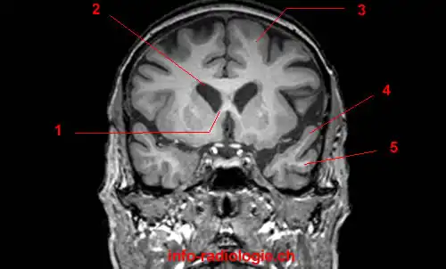
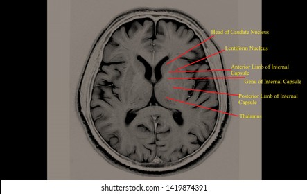
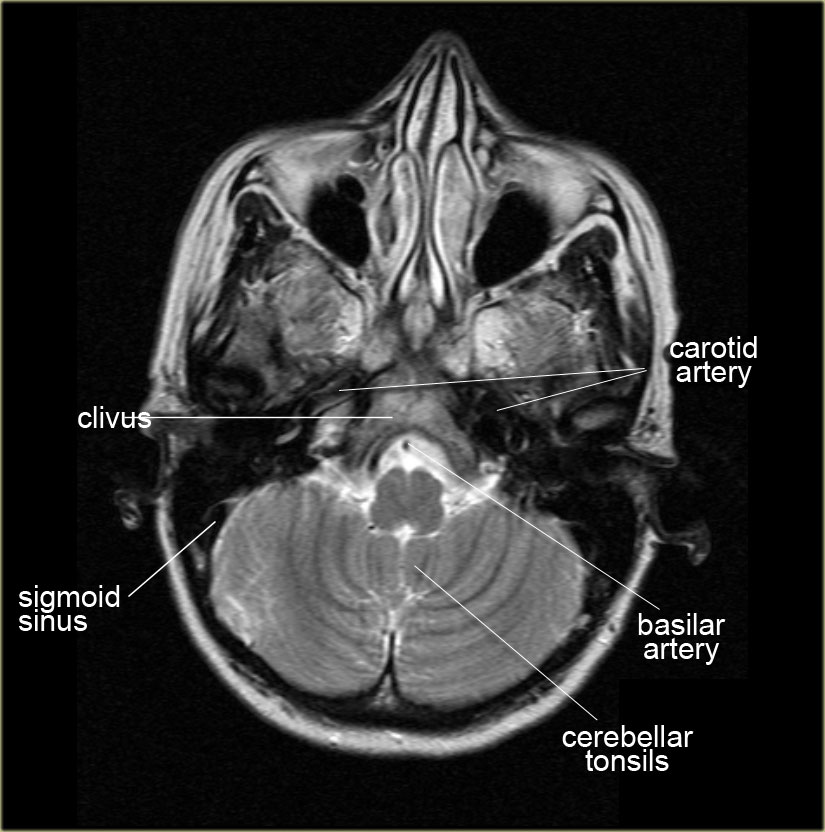
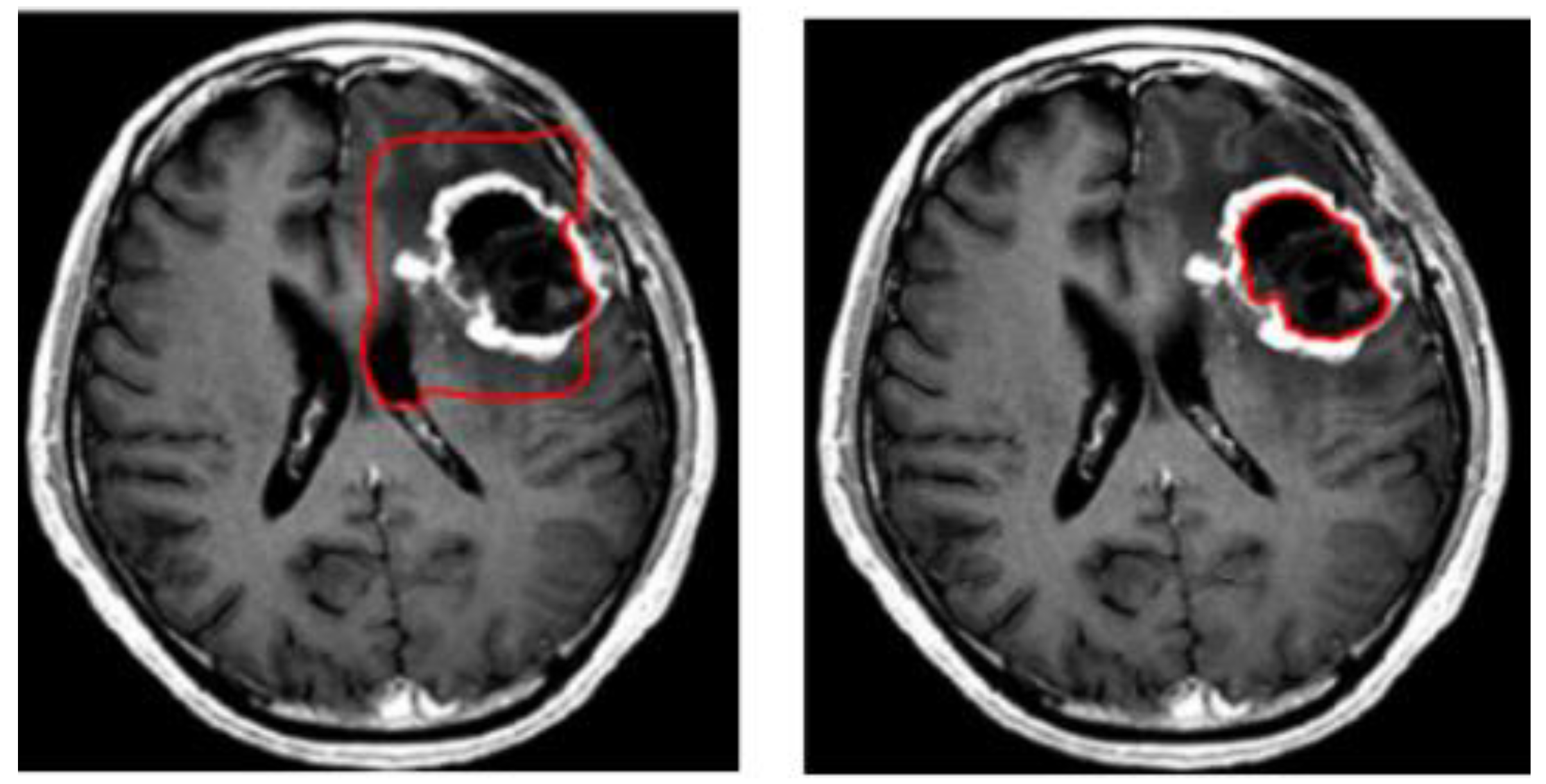
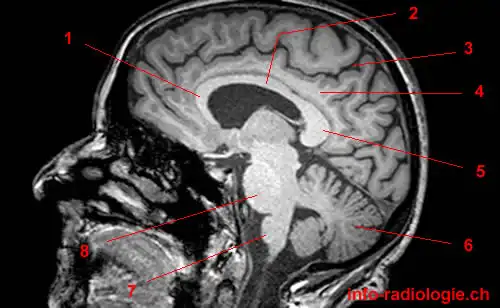
44 brain mri with labels
[2106.09812] Deep reinforcement learning with automated label ... However, labeling images is time-consuming and tedious. We have recently demonstrated that reinforcement learning (RL) can classify 2D slices of MRI brain images with high accuracy. Here we make two important steps toward speeding image classification: Firstly, we automatically extract class labels from the clinical reports. Brain: Atlas of human anatomy with MRI - e-Anatomy - IMAIOS The module on the anatomy of the brain based on MRI with axial slices was redesigned, having received multiple requests from users for coronal and sagittal slices. The elaboration of this new module, its labeling of more than 524 structures on 379 MRI images in three different views and on 26 anatomical diagrams, took more than 6 months.
The MRI Dataset with labels. | Download Scientific Diagram - ResearchGate The identification and classification of tumors in the human mind from MR images at an early stage play a pivotal role in diagnosis such diseases. This work presents the novel Deep Neural network...
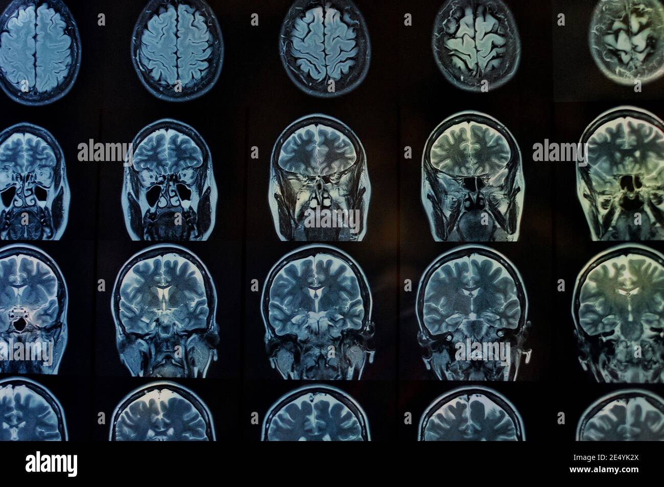
Brain mri with labels
Magnetic Resonance Imaging (MRI) of the Spine and Brain Magnetic resonance imaging (MRI) is a diagnostic procedure that uses a combination of a large magnet, radiofrequencies, and a computer to produce detailed images of organs and structures within the body. Unlike X-rays or computed tomography (CT scans), MRI does not use ionizing radiation. Some MRI machines look like narrow tunnels, while others ... MNI space average T1-weighted MRI of the brain with anatomical labels. MNI space average brain with associated labels. Many of these labels are lifted from Chris Rorden's micron. Efficient Multi-class Fetal Brain Segmentation in High Resolution MRI ... 2.1 Image Acquisition. Multiple low-resolution orthogonal MR sequences of the brain were acquired on 1.5T and 3T clinical GE whole-body scanners at the University Children's Hospital Zurich using a T2-weighted single-shot fast spin echo sequence (ssFSE), with an in-plane resolution of 0.5 × 0.5 mm and a slice thickness of 3-5 mm for 15 subjects.
Brain mri with labels. brain mri with labels | جدني brain mri with contrasts: Transverse Occipital sulcus: brain mri at: how to read a brain mri images: normal mri brain scan image: internal capsule in MRI: ct head brain: brain images mri: mri middle cerebral artery: normal brain image with contrast: brain mri choroid: choroid plexus mri: mri brain normal how to read: occipital foramen MRI: Internal capsule: mri cerebrum Labels · ryx2/MRI-Brain-Registration · GitHub My master's project. Contribute to ryx2/MRI-Brain-Registration development by creating an account on GitHub. 101 labeled brain images and a consistent human cortical labeling protocol In this article we introduce this dataset of manually edited brain image labels applied to the T1-weighted MR images of publicly available multi-modal data acquired from healthy individuals. We also introduce a benchmark for the evaluation of automated registration/segmentation/labeling methods by comparing the manual labels according to this "Desikan-Killiany-Tourville" (DKT) protocol with automatically generated labels. 101 labeled brain images and a consistent human cortical labeling ... given how difficult it is to label brains, the mindboggle-101 dataset is intended to serve as brain atlases for use in labeling other brains, as a normative dataset to establish morphometric variation in a healthy population for comparison against clinical populations, and contribute to the development, training, testing, and evaluation of …
Brain MRI: How to read MRI brain scan | Kenhub MRI is the most sensitive imaging method when it comes to examining the structure of the brain and spinal cord. It works by exciting the tissue hydrogen protons, which in turn emit electromagnetic signals back to the MRI machine. The MRI machine detects their intensity and translates it into a gray-scale MRI image. Labeled MRI Brain Scans - Neuromorphometrics We can also label scans that you provide and we are very interested in labeling white matter anatomy as seen in diffusion-weighted MRI scans. If you want an aggregate version of our data, we can provide it as a probabilistic atlas. The cost to label a single scan is $2449 (USD). Enhancing the REMBRANDT MRI collection with expert segmentation labels ... Pyradiomics 41, an open-source python package was used to extract radiomics features from the segmented labels of the MRI brain scans. It included a total of 120 features, ... Efficient Multi-class Fetal Brain Segmentation in High Resolution MRI ... This work proposes using transfer learning with noisy multi-class labels to automatically segment high resolution fetal brain MRIs using a single set of seg-mentations created with one reconstruction method and tested for generalizability across other reconstruction methods. Segmentation of the developing fetal brain is an important step in quantitative analyses. However, manual segmentation ...
Brain MRI Segmentation with Label Propagation | Semantic Scholar A novel MRI segmentation method is proposed for precisely recognizing main parts of a brain structure and the probable lesion regions based on label propagation mechanism, which is a semi-supervised learning method in machine learning literatures. Volume 2, Issue 5 September - October 2013 Page 158 Abstract: Magnetic Resonance Imaging (MRI) is a widely used method of medical imaging to study ... Similarity Measure-Based Possibilistic FCM With Label Information for ... Magnetic resonance imaging (MRI) is extensively applied in clinical practice. Segmentation of the MRI brain image is significant to the detection of brain abnormalities. However, owing to the coexistence of intensity inhomogeneity and noise, dividing the MRI brain image into different clusters precisely has become an arduous task. In this paper, an improved possibilistic fuzzy c-means (FCM ... MRI anatomy | free MRI axial brain anatomy - Mrimaster.com This MRI brain cross sectional anatomy tool is absolutely free to use. Use the mouse scroll wheel to move the images up and down alternatively use the tiny arrows (>>) on both side of the image to move the images. Label all the features of brain MRI after Image Segmentation? I want to label the features of brain MRI such as fluid, midline etc. in MATLAB after segmentation, Image is segmented with Canny algorithm for better results , any idea please.
Labeled imaging anatomy cases | Radiology Reference Article ... CT head: non-contrast coronal. CT head: non-contrast sagittal. CT head: angiogram axial. CT head: angiogram coronal. CT head: angiogram sagittal. CT head: venogram axial. CT head: venogram coronal. CT head: venogram sagittal. CT head: venogram sagittal (diagram)
Are there any publicly available datasets for brain MRI images with labels? The human neocortex (the outer part of the brain) has roughly 10^4-10^5 neurons per cubic millimeter ( ). However that is at the very extreme, and is only for structural MRI. For functional MRI, the data are acquired at a resolution of about 0.5*0.5*1.0 mm, giving a resolution of about 2500-25000 neuro
Arterial spin labeling MRI: clinical applications in the brain Arterial spin labeling (ASL) perfusion magnetic resonance imaging (MRI) sequences are increasingly being used to provide MR-based CBF quantification without the need for contrast adm … Visualization of cerebral blood flow (CBF) has become an important part of neuroimaging for a wide range of diseases.
Brain MRI: What It Is, Purpose, Procedure & Results - Cleveland Clinic A brain MRI (magnetic resonance imaging) scan, also called a head MRI, is a painless procedure that produces very clear images of the structures inside of your head — mainly, your brain. MRI uses a large magnet, radio waves and a computer to produce these detailed images. It doesn't use radiation. Currently, MRI is the most sensitive imaging test ...
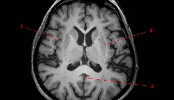
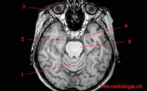
Cross-sectional anatomy of the brain - e-Anatomy - IMAIOS Axial MRI Atlas of the Brain. Free online atlas with a comprehensive series of T1, contrast-enhanced T1, T2, T2*, FLAIR, Diffusion -weighted axial images from a normal humain brain. Scroll through the images with detailed labeling using our interactive interface. Perfect for clinicians, radiologists and residents reading brain MRI studies.
normal mri brain with labels | جدني فيما يلي صفحات متعلقة بكلمة البحث: normal mri brain with labels. Brain MRI. 06/03/2012 11:47:00 ص ...
Brain MRI segmentation | Kaggle This dataset contains brain MR images together with manual FLAIR abnormality segmentation masks. The images were obtained from The Cancer Imaging Archive (TCIA). They correspond to 110 patients included in The Cancer Genome Atlas (TCGA) lower-grade glioma collection with at least fluid-attenuated inversion recovery (FLAIR) sequence and genomic cluster data available.
Brain MRI Images for Brain Tumor Detection | Kaggle Brain MRI Images for Brain Tumor Detection. Data. Code (249) Discussion (8)
Brain lobes - annotated MRI | Radiology Case | Radiopaedia.org Neuro- MRI by Dr Bálint Botz Head MRI by Anthony Pennuto; Annotated CT/MR Teaching by Matt Wong Brain Anatomy MRI by R. Furman Borst MD; fälle für Anatomie by Eva Fischer NRAD by Johann Jende; Частки ГМ 2021 by Василь; Annotated Anatomy by Marc Hidalgo; 6_NEUROLOGIC IMAGING - Weissleder by Felicia Wright; MRI BRAIN by Dr Mohammed Sharikh
Classification of brain tumours in MR images using deep ... - Nature The healthy brain class achieved the highest F1 score of 0.9998 \({\pm }\) 0.0002, with the pre-trained ResNet 3D model, which can be expected because of the complete absence of any lesion in the ...
Atlas of BRAIN MRI - W-Radiology Brain magnetic resonance imaging (MRI) is a common medical imaging method that allows clinicians to examine the brain's anatomy (1). It uses a magnetic field and radio waves to produce detailed images of the brain and the brainstem to detect various conditions (2).
Efficient Multi-class Fetal Brain Segmentation in High Resolution MRI ... 2.1 Image Acquisition. Multiple low-resolution orthogonal MR sequences of the brain were acquired on 1.5T and 3T clinical GE whole-body scanners at the University Children's Hospital Zurich using a T2-weighted single-shot fast spin echo sequence (ssFSE), with an in-plane resolution of 0.5 × 0.5 mm and a slice thickness of 3-5 mm for 15 subjects.
MNI space average T1-weighted MRI of the brain with anatomical labels. MNI space average brain with associated labels. Many of these labels are lifted from Chris Rorden's micron.
Magnetic Resonance Imaging (MRI) of the Spine and Brain Magnetic resonance imaging (MRI) is a diagnostic procedure that uses a combination of a large magnet, radiofrequencies, and a computer to produce detailed images of organs and structures within the body. Unlike X-rays or computed tomography (CT scans), MRI does not use ionizing radiation. Some MRI machines look like narrow tunnels, while others ...
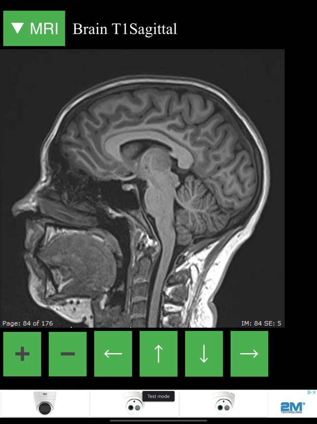

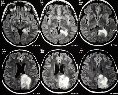


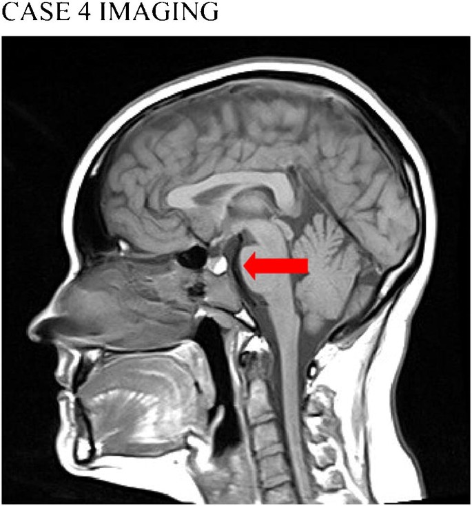









:watermark(/images/watermark_only_sm.png,0,0,0):watermark(/images/logo_url_sm.png,-10,-10,0):format(jpeg)/images/anatomy_term/thalamus-12/JikayR0budNwnh0ynTMMA_Thalamus.jpg)




Post a Comment for "44 brain mri with labels"