39 microscope diagram with labels and definitions
Compound Microscope Parts, Functions, and Labeled Diagram Compound Microscope Parts, Functions, and Labeled Diagram Parts of a Compound Microscope Each part of the compound microscope serves its own unique function, with each being important to the function of the scope as a whole. Simple Microscope - Definition, Types, Working Principle & Formula Compound Microscope. 1. Simple microscope comprises a biconvex lens used as a magnifying glass. Compound microscope comprises 2 or more convex lenses where one lens is the eyepiece and the other one is the objective lens. 2. Natural light is the source to see the object. An illuminator is a source to see the object. 3.
Fuzzy concept - Wikipedia A fuzzy concept is a concept of which the boundaries of application can vary considerably according to context or conditions, instead of being fixed once and for all. This means the concept is vague in some way, lacking a fixed, precise meaning, without however being unclear or meaningless altogether. It has a definite meaning, which can be made more precise only …

Microscope diagram with labels and definitions
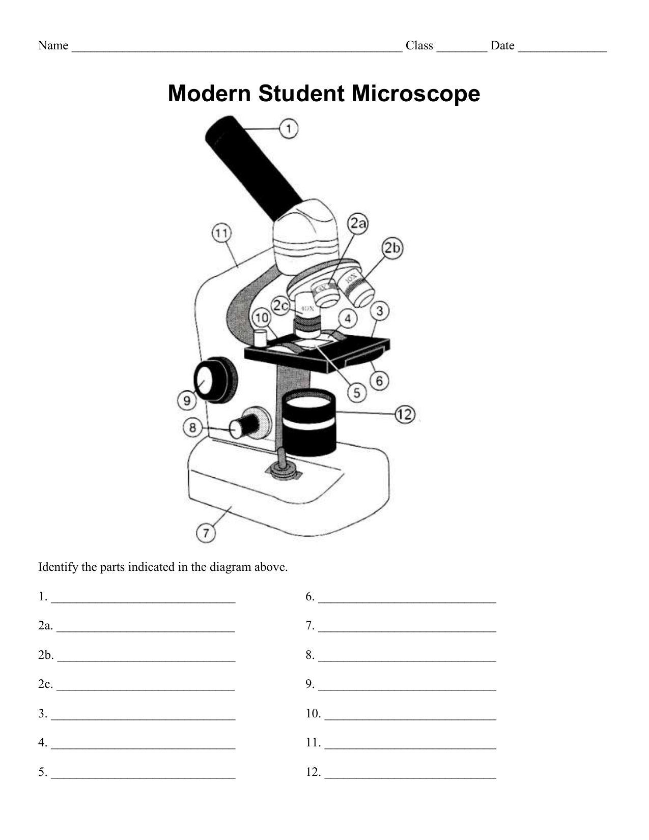
Parts of a microscope with functions and labeled diagram Microscope Definition Microscopes are instruments that are used in science laboratories to visualize very minute objects such as cells, and microorganisms, giving a contrasting image that is magnified. Microscopes are made up of lenses for magnification, each with its own magnification powers. 16.3 Lenses - Physics | OpenStax Figure 16.26 Rays of light enter a concave, or diverging, lens parallel to its axis diverge and thus appear to originate from its focal point, F. The dashed lines are not rays; they indicate the directions from which the rays appear to come. The focal length, ƒ, of a diverging lens is negative.An expanded view of the path taken by ray 1 shows the perpendiculars and the … PDF Parts of a Microscope Printables - Homeschool Creations Label the parts of the microscope. You can use the word bank below to fill in the blanks or cut and paste the words at the bottom. Microscope Created by Jolanthe @ HomeschoolCreations.net. Parts of a eyepiece arm stageclips nosepiece focusing knobs illuminator stage objective lenses
Microscope diagram with labels and definitions. Simple Microscope - Diagram (Parts labelled), Principle, Formula and Uses A simple microscope consists of Optical parts Mechanical parts Labeled Diagram of simple microscope parts Optical parts The optical parts of a simple microscope include Lens Mirror Eyepiece Lens A simple microscope uses biconvex lens to magnify the image of a specimen under focus. Parts of Stereo Microscope (Dissecting microscope) - labeled diagram ... Labeled part diagram of a stereo microscope Major structural parts of a stereo microscope Optical components of a stereo microscope - definition and function Eyepieces Eyepiece tube Diopter adjustment ring Interpupillary Adjustment Objective Lenses Barlow lens Adjustment Knobs Light sources Stage plate Stage chips Compound Microscope - Diagram (Parts labelled), Principle and Uses See: Labeled Diagram showing differences between compound and simple microscope parts Structural Components The three structural components include 1. Head This is the upper part of the microscope that houses the optical parts 2. Arm This part connects the head with the base and provides stability to the microscope. Metalloid - Wikipedia Definitions. Judgment-based. A metalloid is an element that possesses a preponderance of properties in between, or that are a mixture of, those of metals and nonmetals, and which is therefore hard to classify as either a metal or a nonmetal. This is a generic definition that draws on metalloid attributes consistently cited in the literature. Difficulty of categorisation is a key …
Parts of the Microscope with Labeling (also Free Printouts) Parts of the Microscope with Labeling (also Free Printouts) A microscope is one of the invaluable tools in the laboratory setting. It is used to observe things that cannot be seen by the naked eye. Table of Contents 1. Eyepiece 2. Body tube/Head 3. Turret/Nose piece 4. Objective lenses 5. Knobs (fine and coarse) 6. Stage and stage clips 7. Aperture Compound Microscope Parts, Diagram Definition, Application, Working ... Compound Microscope Parts, Diagram Definition, Application, Working Principle. ... For future reference, adhesive labels are stuck to the base and sides of the microscope. (iii) Use. After calibrating the eyepiece scales for all objective lenses, the microscope can be used to measure the dimensions and morphology of cells and sub-cellular ... Microscope Parts & Functions - AmScope Microscope Terms. This is a glossary of commonly used microscopy terms. Abbe Condenser: A lens that is specially designed to mount under the stage and which typically moves in a vertical direction. An adjustable iris controls the diameter of the beam of light entering the lens system. Both by changing the size of this iris and by moving the ... Simple Microscope - Parts, Functions, Diagram and Labelling Simple Microscope - Parts, Functions, Diagram and Labelling A microscope is one of the commonly used equipment in a laboratory setting. A microscope is an optical instrument used to magnify an image of a tiny object; objects that are not visible to the human eyes. Table of Contents The common types of microscopes are: What is a Simple microscope?
› books › NBK310485Laboratory procedures for diagnosis of anthrax, and isolation ... 1. Anthrax and the microbiology laboratory; operational safety. With some country-to-country variation in safety level definitions and requirements, recommendations for the manipulation of the causative agent of anthrax, Bacillus anthracis, generally are that BSL (biosafety level) 2 practices, containment equipment and facilities are appropriate for diagnostic tests, but BSL3 standards should ... PDF Definitions of the Parts of the Microscope - ualberta.ca microscope moves the stage up and down to bring the specimen into focus. The gearing mechanism of the adjustment produces a large vertical movement of the stage with only a partial revolution of the knob. Because of this, the coarse adjustment should only be used with low power (4X and 10X objectives) and never with the high power lenses (40X and Laboratory procedures for diagnosis of anthrax, and isolation and ... 1. Anthrax and the microbiology laboratory; operational safety. With some country-to-country variation in safety level definitions and requirements, recommendations for the manipulation of the causative agent of anthrax, Bacillus anthracis, generally are that BSL (biosafety level) 2 practices, containment equipment and facilities are appropriate for diagnostic tests, but BSL3 … Microscope Parts and Functions With Labeled Diagram and Functions How ... First, the purpose of a microscope is to magnify a small object or to magnify the fine details of a larger object in order to examine minute specimens that cannot be seen by the naked eye. Here are the important compound microscope parts... Eyepiece: The lens the viewer looks through to see the specimen.
openstax.org › books › physics16.3 Lenses - Physics | OpenStax The ray diagram in Figure 16.33 shows image formation by the cornea and lens of the eye. The rays bend according to the refractive indices provided in Table 16.4 . The cornea provides about two-thirds of the magnification of the eye because the speed of light changes considerably while traveling from air into the cornea.
16 Parts of a Compound Microscope: Diagrams and Video Body of the Microscope In compound microscopes with two eye pieces there are prisms contained in the body that will also split the beam of light to enable you to view the image through both eye pieces. 2. Arm The arm of the microscope is another structural piece. The arm connects the base of the microscope to the head/body of the microscope.
› 44090147 › CambridgeCambridge International AS and A Level Biology Coursebook ... Enter the email address you signed up with and we'll email you a reset link.
microscope labels Flashcards | Quizlet Start studying microscope labels. Learn vocabulary, terms, and more with flashcards, games, and other study tools.

Animal Cell Cake With Labels - Edible Cell Project Chocolate Chip Cookie Science Hip Homeschool ...
Mapping Genomes - Genomes - NCBI Bookshelf Draw a diagram showing how a double-stranded cDNA is synthesized. 15. Define the term ‘mapping reagent’ and explain how a panel of radiation hybrids is used as a mapping reagent. 16. Explain how a clone library is used as a mapping reagent. 17. Draw a diagram to show how a sample of a single human chromosome can be obtained by flow cytometry.
The Parts of a Microscope (Labeled) Printable - TeacherVision This diagram labels and explains the function of each part of a microscope. Use this printable as a handout or transparency to help prepare students for working with laboratory equipment. ... The Parts of a Microscope (Labeled) Printable. Download. Add to Favorites. CREATE NEW FOLDER. Cancel. Manage My Favorites. Share. This diagram labels and ...
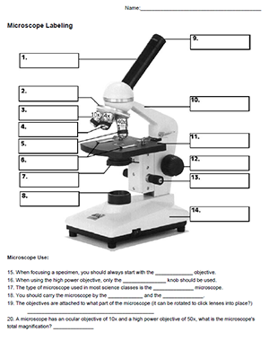
Label the microscope — Science Learning Hub In this interactive, you can label the different parts of a microscope. Use this with the Microscope parts activity to help students identify and label the main parts of a microscope and then describe their functions. Drag and drop the text labels onto the microscope diagram.
Compound Microscope: Definition, Diagram, Parts, Uses, Working ... - BYJUS A compound microscope is defined as. A microscope with a high resolution and uses two sets of lenses providing a 2-dimensional image of the sample. The term compound refers to the usage of more than one lens in the microscope. Also, the compound microscope is one of the types of optical microscopes. The other type of optical microscope is a ...
Compound Microscope- Definition, Labeled Diagram, Principle, Parts, Uses The term "compound" in compound microscopes refers to the microscope having more than one lens. Devised with a system of combination of lenses, a compound microscope consists of two optical parts, namely the objective lens and the ocular lens. Working Principle of the Compound Microscope
A Study of the Microscope and its Functions With a Labeled Diagram To better understand the structure and function of a microscope, we need to take a look at the labeled microscope diagrams of the compound and electron microscope. These diagrams clearly explain the functioning of the microscopes along with their respective parts. Man's curiosity has led to great inventions. The microscope is one of them.
Fountain Essays - Your grades could look better! All our academic papers are written from scratch. All our clients are privileged to have all their academic papers written from scratch. These papers are also written according to your lecturer’s instructions and thus minimizing any chances of plagiarism.
E-Learning – AOAC India Bright Field Microscope, Dark Field Microscope, Phase Contrast Microscope, Fluorescence Microscope, Confocal microscopy, Scanning and Transmission Electron Microscope and applications: 17th June 2019, 2019 11.30-12.30 pm: Watch Video: 50 : Nuclear magnetic resonance (NMR) – Part 1 DR. CHANDRASHEKHAR MR. RAGHAV MAVINKURVE, BRUKER
Compound Microscope Parts - Labeled Diagram and their Functions - Rs ... The eyepiece (or ocular lens) is the lens part at the top of a microscope that the viewer looks through. The standard eyepiece has a magnification of 10x. You may exchange with an optional eyepiece ranging from 5x - 30x. [In this figure] The structure inside an eyepiece. The current design of the eyepiece is no longer a single convex lens.
Microscope, Microscope Parts, Labeled Diagram, and Functions Microscope, Microscope Parts, Labeled Diagram, and Functions What is Microscope? A microscope is a laboratory instrument used to examine objects that are too small to be seen by the naked eye. It is derived from Ancient Greek words and composed of mikrós, "small" and skopeîn,"to look" or "see".
CODEX multiplexed tissue imaging with DNA-conjugated ... - Nature 02.07.2021 · This protocol describes co-detection by indexing, a highly multiplexed imaging technology that uses DNA-conjugated antibodies to image up to 60 markers in formalin-fixed, paraffin-embedded and ...
› articles › s41596/021/00556-8CODEX multiplexed tissue imaging with DNA-conjugated ... - Nature Jul 02, 2021 · A newly adapted version of CODEX uses an automated microfluidics system and conventional fluorescent microscope to iteratively hybridize, image and strip fluorescently labeled DNA probes that are ...


Post a Comment for "39 microscope diagram with labels and definitions"