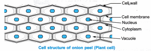
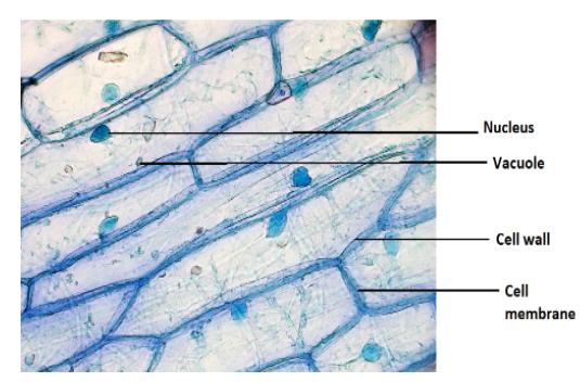
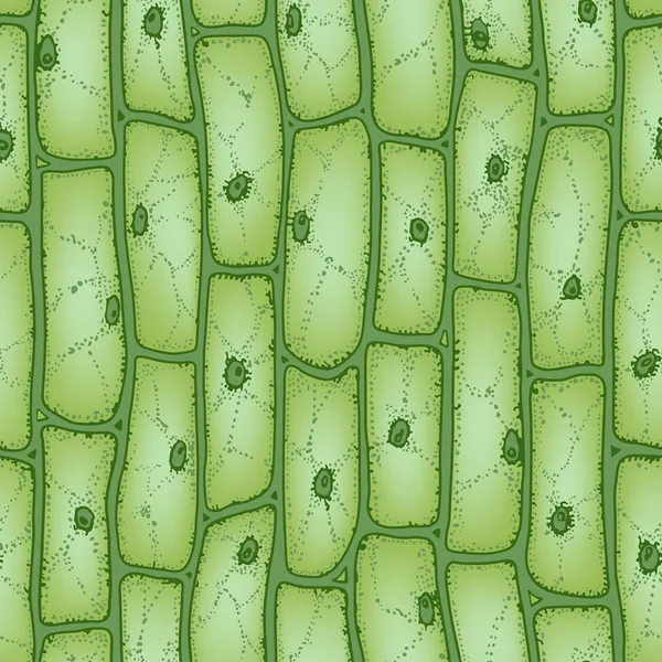
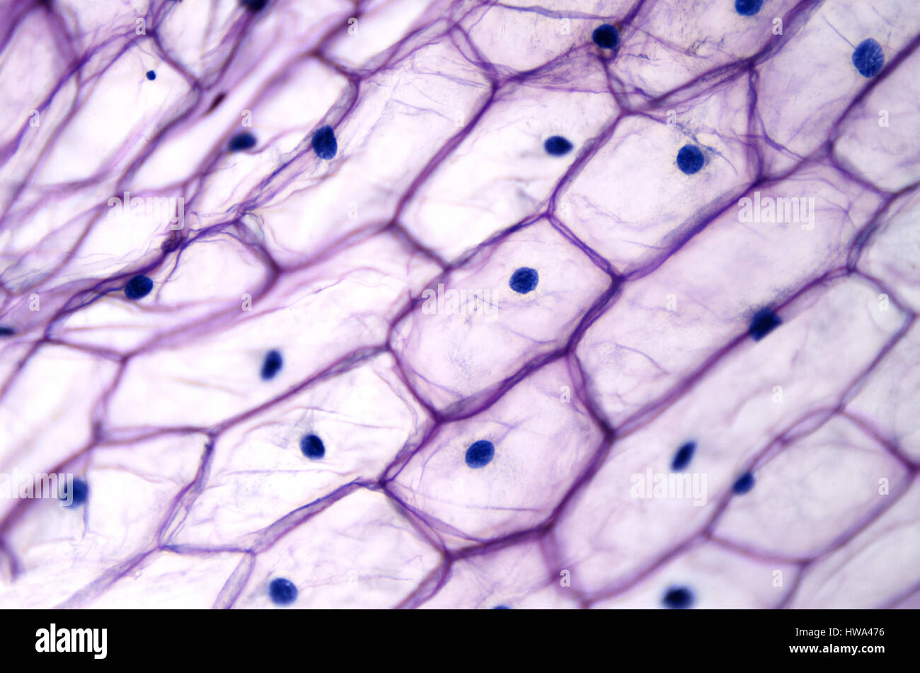
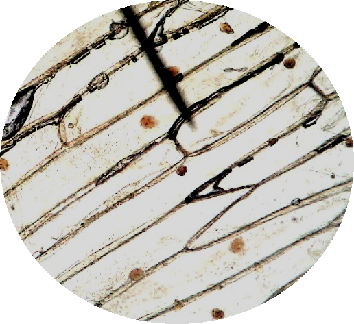
38 onion cells under microscope with labels
How to observe onion cells under a microscope? - JacAnswers How to observe onion cells under a microscope? Gently lay a microscopic cover slip on the membrane and press it down gently using a needle to remove air bubbles. Touch a blotting paper on one side of the slide to drain excess iodine/water solution, Place the slide on the microscope stage under low power to observe. Adjust focus for clarity to ... onion cells under a microscope labeled Onion epidermal cells, iodine stain, 400X. distichum 'Spence' (C and D), and sorghum cv BTx623 (E and F) grown in freshwater were imaged under a light microscope Location Tiruvanmiyur, Chennai 600041 Financial Sentiment Analysis Github Label an de diagram of a stomatal apparatus 2 four parts on the diagram Inference The cells observed under the ...
› blog › postsLyft's Commitment to Climate Action - Lyft Blog A lot of voters agree with us. Early support for the measure is strong. What started with good policy created by a diverse group of organizations — including the Natural Resources Defense Council, the American Lung Association, California State Firefighters, the Coalition for Clean Air, the State Association of Electrical Workers – IBEW, the San Francisco Bay Area Planning and Urban ...
Onion cells under microscope with labels
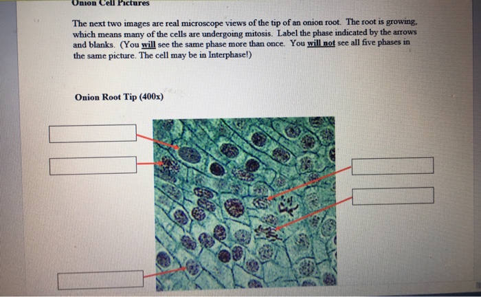
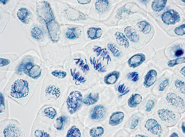
Structure and function of mitochondrial membrane protein … Oct 29, 2015 · Mitochondria can be seen in the light microscope, but their detailed internal structure is only revealed by electron microscopy. In the 1990s, the structure of mitochondria was investigated by electron tomography of thin plastic sections [].While this yielded striking three-dimensional (3D) images of their internal membrane system, molecular detail was lost due to … Onion Root Tip Mitosis - Stages, Experiment and Results - MicroscopeMaster · Place a cap/lid onto the vial (ensure that the cap/lid has a pinprick hole) and place the vial in the water bath (at 55 degrees C) for about 5 minutes - This enhances the staining process · Using the forceps, remove the root tips from the vial of stain and place them onto a clean microscope glass slide Lyft's Commitment to Climate Action - Lyft Blog A lot of voters agree with us. Early support for the measure is strong. What started with good policy created by a diverse group of organizations — including the Natural Resources Defense Council, the American Lung Association, California State Firefighters, the Coalition for Clean Air, the State Association of Electrical Workers – IBEW, the San Francisco Bay Area Planning and …
Onion cells under microscope with labels. bmcbiol.biomedcentral.com › articles › 10Structure and function of mitochondrial membrane protein ... Oct 29, 2015 · Biological energy conversion in mitochondria is carried out by the membrane protein complexes of the respiratory chain and the mitochondrial ATP synthase in the inner membrane cristae. Recent advances in electron cryomicroscopy have made possible new insights into the structural and functional arrangement of these complexes in the membrane, and how they change with age. This review places ... Vacuole Function and Structure – Extra Space Storage How to see the vacuole under a microscope. ... An example of using Neutral red to stain fresh onion cells. (A) Neutral red stains vacuoles only in viable cells. (B,C) When cells are damaged by high pressure, cell integrity loses, and vacuoles leak. ... BCECF is a chemical that labels the acidic lumen of the vacuole. FM4-64 and MDY-64 can label ... What do you observe onion cells under a microscope? Staining Onion Cells. Since onion peels are translucent, you'll need to stain the onion cells before you observe them under the microscope. There are different types of stains depending on what type of cell you are going to look at. Iodine- dark stain that colors starches in cells. In an onion cell, it will make the cell wall more visible. Worksheets Index - The Biology Corner CELLS Cell Parts. Cheek Cell Lab - observe cheek cells under the microscope Cheek Cell Virtual Lab – if you missed it in class Animal Cell Coloring - color a typical animal cell. Plant Cell Coloring - color a typical plant cell Plant Cell Lab - microscope observation of onion and elodea Plant Cell Lab Makeup - microscope observation of onion and elodea, if students missed the …
Onion Cells Under Microscope With Labels - Realtec Find and download Onion Cells Under Microscope With Labels image, wallpaper and background for your Iphone, Android or PC Desktop. Realtec have about 34 image published on this page. onion microscope under cells cepa allium slide footage shutterstock background royalty Pin It Share Download Alopecia Areata: Causes, Symptoms, and Diagnosis - Healthline Mar 01, 2022 · Alopecia areata is an autoimmune condition.An autoimmune condition develops when the immune system mistakes healthy cells for foreign substances. Normally, the immune system defends your body ... Cambridge Lower Secondary Science Learner's Book 7 sample Oct 13, 2020 · Try not to get air bubbles under the cover slip. 6 Turn the objective lenses on the microscope until the smallest one is over the hole in the stage. ... • I saw onion cells down the microscope ... Onion Skin Epidermis Sample under microscope 4x,10x Magnification A sample on an onion skin epidermis diyed in blue for visibility, viewd under the microscope at 4x and 10x magnification.microscope:Biolux model :AL
How to see onion cell with Homemade Microscope Easy - YouTube Hi friends I am Khilesh and going to show you onion cells in my homemade microscope. Tip:- Stain the cells with water then with iodine then again with water.... Observing Onion Cells Under The Microscope » Microscope Club Afterwards, carefully mount the prepared and stained onion cell slide onto the microscope stage. Make sure that the cover slip is perfectly aligned with the microscope slide, and that any excess stain has been wiped off. Secure the slide on the stage using the stage clips. Onion Plant Cell Under Microscope Labeled / Onion Cells - Onion ... Onion Plant Cell Under Microscope Labeled / Onion Cells - Onion epidermis with pigmented large cells.. Under the microscope, animal cells appear different based on the type of the cell. Human cheek cells and to record observations and draw their labelled diagrams. However, it is too small to see through a microscope. Microsoft takes the gloves off as it battles Sony for its Activision ... Oct 12, 2022 · Microsoft pleaded for its deal on the day of the Phase 2 decision last month, but now the gloves are well and truly off. Microsoft describes the CMA’s concerns as “misplaced” and says that ...
About Our Coalition - Clean Air California About Our Coalition. Prop 30 is supported by a coalition including CalFire Firefighters, the American Lung Association, environmental organizations, electrical workers and businesses that want to improve California’s air quality by fighting and preventing wildfires and reducing air pollution from vehicles.
yeson30.org › aboutAbout Our Coalition - Clean Air California About Our Coalition. Prop 30 is supported by a coalition including CalFire Firefighters, the American Lung Association, environmental organizations, electrical workers and businesses that want to improve California’s air quality by fighting and preventing wildfires and reducing air pollution from vehicles.
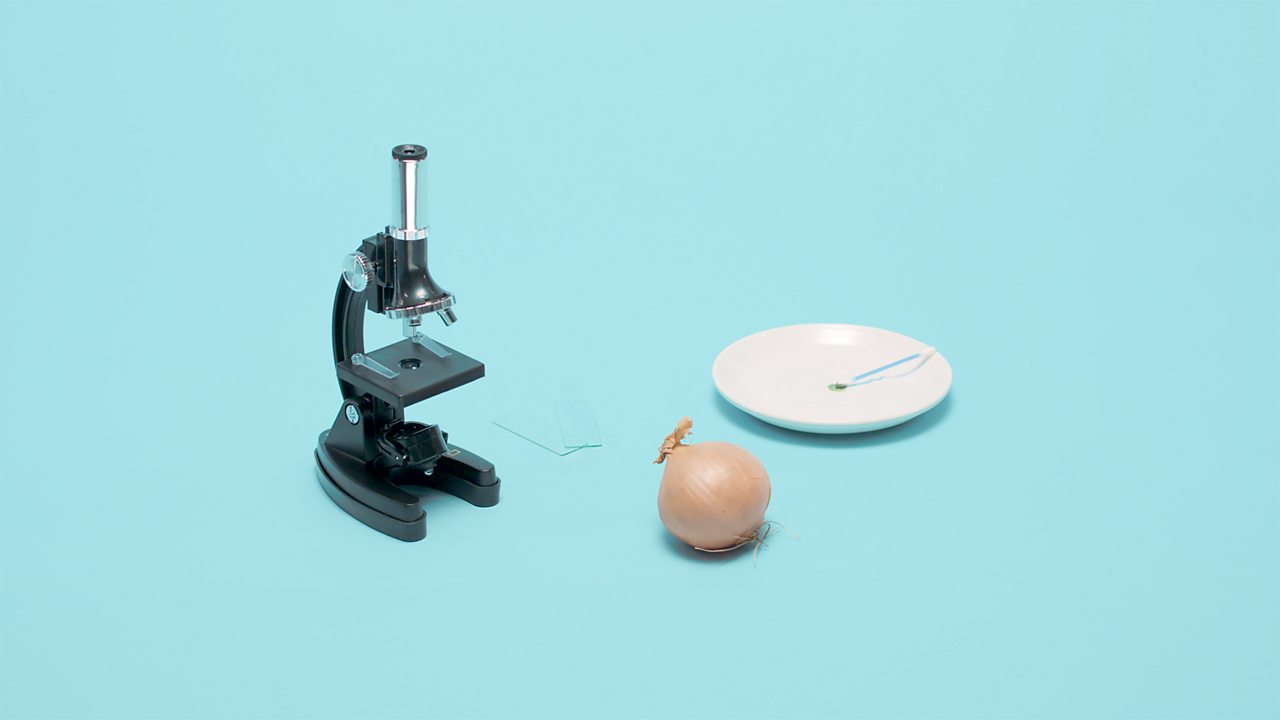
Microscopy, size and magnification (CCEA) - BBC Bitesize cells from the inside of an onion. Place cells on a microscope slide. Add a drop of water or iodine (a chemical stain). Lower a coverslip onto the onion cells using forceps or a mounted needle.
The Cell Structure of an Onion | Sciencing Onion cells are among the most common choices for cell studies in early biology classes. Easily obtained, inexpensive, they offer samples with no difficult technique required. The thin layer of skin found on the inside of an onion scale (one layer of onion) lifts off without effort and can be wet mounted on a slide with no need for extreme skill.
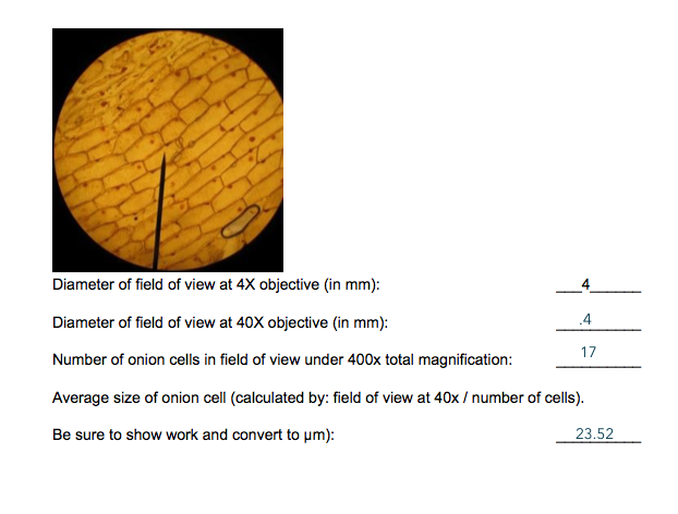
PDF Onion Cell Lab Research Biology Onion Cell Lab page 1 of 3 Onion Cell Lab After you have completed the rest of this lab come back to this cover page DRAW & LABEL AN ONION CELL WITH ALL THE PARTS / ORGANELLES YOU OBSERVE UNDER 40X. Purpose: To observe and identify major plant cell structures and to relate the structure of the cell to its function. Materials: 1 ...
PPIC Statewide Survey: Californians and Their Government Oct 27, 2022 · Key Findings. California voters have now received their mail ballots, and the November 8 general election has entered its final stage. Amid rising prices and economic uncertainty—as well as deep partisan divisions over social and political issues—Californians are processing a great deal of information to help them choose state constitutional officers and …
Onion Cell Lab Report.docx - Onion Cell Lab Report By Onion Cell Lab Report By : Nawaf Almalki Introduction: Many things that are viewed using a microscope, particularly cells, can appear quite transparent under the microscope. The internal parts of the cells, the organelles, are so transparent that they are often difficult to see. Biologists have developed a number of stains that help them see the cells and their organelles by adding color to ...
onion cells under a microscope labeled - sevadham.net Onion Cells Under a Microscope Requirements Preparation and Observation. Gently lay a microscopic cover slip on the membrane and press it down gently using a needle to remove air The diagram is very clear and labeled. In this video you will see onion cells under a microscope (100x-2500x) as is, without any coloring.
issuu.com › cupeducation › docsCambridge Lower Secondary Science Learner's Book 7 sample - Issuu Oct 13, 2020 · Try not to get air bubbles under the cover slip. 6 Turn the objective lenses on the microscope until the smallest one is over the hole in the stage. ... • I saw onion cells down the microscope ...
DOC Plant and Animal Cells Microscope Lab - hillsboro.k12.oh.us Students will observe onion cells under a microscope. Students will discover that their skin is made up of cells. Students will observe cheek cells under a microscope. Materials: microscope. ... Draw a diagram of one cheek cell and label the parts. (You should observe the cell membrane, nucleus, and cytoplasm.)
Under the Micrsocope: Onion Cell (100x - 400x) - YouTube In this "experiment" we will see onion cells under the microscope.For the experiment you will only need onion, dropper and the microscope (container and tool...
U.S. appeals court says CFPB funding is unconstitutional - Protocol Oct 20, 2022 · That battle could introduce significant uncertainty for the many fintech businesses that fall under the agency’s purview. The decision. A three-judge panel of the New Orleans-based 5th Circuit Court of Appeals found Wednesday that the CFPB’s funding structure violated the Constitution’s separation of powers doctrine.
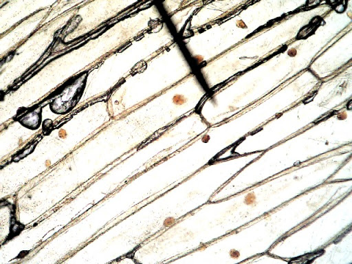
What organelles are in an onion cell? - Biology Stack Exchange You cannot see most of these as they appear translucent as well as being too small to see under the light microscope. You need an electron microscope to view these. ... chloroplasts are not present in an onion cell as it is not a photosynthesising cell. This is a typical onion cell slide with labels: Share. Improve this answer. Follow edited ...
› 2022/10/12 › 23400986Microsoft takes the gloves off as it battles Sony for its ... Oct 12, 2022 · Microsoft pleaded for its deal on the day of the Phase 2 decision last month, but now the gloves are well and truly off. Microsoft describes the CMA’s concerns as “misplaced” and says that ...
rsscience.com › vacuole-function-and-structureVacuole Function and Structure - Extra Space Storage - Rs ... How to see the vacuole under a microscope. Although the vacuole does not take as much dye as other organelles of the cell (the vacuole does not contain many stainable constituents), you can still see and study the structure of vacuoles under a compound microscope. The trick is to use dyes that can stain the cell sap inside the vacuole.
› worksheetsWorksheets Index - The Biology Corner Plant Cell Lab Makeup - microscope observation of onion and elodea, if students missed the lab that day they can view a site with pictures to complete lab handout Plant Cell Virtual Lab – use a virtual microscope to view plant cells. Comparing Plant and Animal Cells – looks at cheek and onion cells
Onion Cells Under a Microscope - Requirements/Preparation/Observation Add a drop of iodine solution on the onion membrane (or methylene blue) Gently lay a microscopic cover slip on the membrane and press it down gently using a needle to remove air bubbles. Touch a blotting paper on one side of the slide to drain excess iodine/water solution, Place the slide on the microscope stage under low power to observe.
Microscope Cell Lab: Cheek, Onion, Zebrina | SchoolWorkHelper Microscope Cell Lab: Cheek, Onion,…. Introduction. The purpose of this lab was to use the microscope and identify cells such as animal cells and plant cells. This subject is important because in Biology, we will be using the microscope many times during different laboratory exercises. The microscope is used for looking at many specimens that ...
Lyft's Commitment to Climate Action - Lyft Blog A lot of voters agree with us. Early support for the measure is strong. What started with good policy created by a diverse group of organizations — including the Natural Resources Defense Council, the American Lung Association, California State Firefighters, the Coalition for Clean Air, the State Association of Electrical Workers – IBEW, the San Francisco Bay Area Planning and …
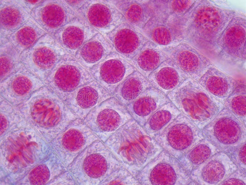
Onion Root Tip Mitosis - Stages, Experiment and Results - MicroscopeMaster · Place a cap/lid onto the vial (ensure that the cap/lid has a pinprick hole) and place the vial in the water bath (at 55 degrees C) for about 5 minutes - This enhances the staining process · Using the forceps, remove the root tips from the vial of stain and place them onto a clean microscope glass slide
Structure and function of mitochondrial membrane protein … Oct 29, 2015 · Mitochondria can be seen in the light microscope, but their detailed internal structure is only revealed by electron microscopy. In the 1990s, the structure of mitochondria was investigated by electron tomography of thin plastic sections [].While this yielded striking three-dimensional (3D) images of their internal membrane system, molecular detail was lost due to …











Post a Comment for "38 onion cells under microscope with labels"