39 respiratory system diagram with labels
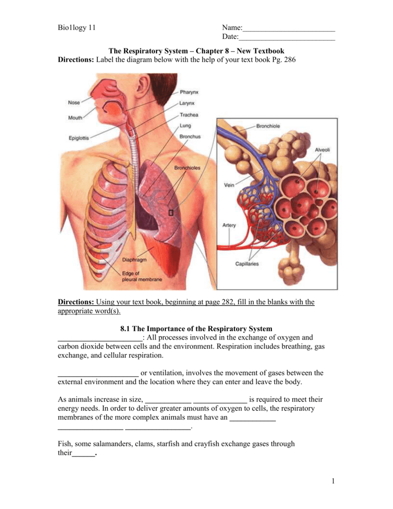
› main › UnitResourceScience A-Z Human Body Grades 3-4 Life Science Unit This unit focuses on the following body systems: skeletal, muscular, nervous, respiratory, circulatory, digestive, and excretory. Overview Resources Print Unit Guide PDF Project Unit Guide Respiratory system: Anatomy and functions | Kenhub The lower respiratory tract includes the larynx below the vocal cords, the trachea, bronchi, bronchioles and the lungs. The lungs are most often considered as part of the lower respiratory tract, but are sometimes described as a separate entity. They contain the respiratory bronchioles, alveolar ducts, alveolar sacs and alveoli .
Respiratory system - Wikipedia The tract is divided into an upper and a lower respiratory tract. The upper tract includes the nose, nasal cavities, sinuses, pharynx and the part of the larynx above the vocal folds. The lower tract (Fig. 2.) includes the lower part of the larynx, the trachea, bronchi, bronchioles and the alveoli .
Respiratory system diagram with labels
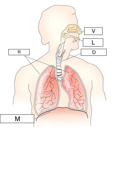
Drawing Of Lungs With Labels / Lung Cancer Icon In Outline Style ... Labeled diagram of the lungs/respiratory system.: A beautiful drawing of lung. Label bronchial tree on any lungs. First by outlining the lungs, bronchi, and trachea, then detailing additional. And it will teach you to draw the lung very easily. Give the diagram a heading and . Lung cancer is a leading cause of death in the u.s. Respiratory System Of Human Body: Anatomy, Parts, and Respiration The human respiratory system consisted of a pair of nostrils openings out above the upper lips. The nasal passage starts from the nostrils and ends in the nasal chamber. This passage opens into the pharynx. It is the common passage for food and air and it is connected with the mouth and nose. This pharynx is connected with the trachea via Larynx. [MCQ] - Carefully study the diagram of the human respiratory system Carefully study the diagram of the human respiratory system with labels A, B, C and D. Select the option which gives correct identification and main function and /or characteristic.(A) (i) Trachea: It is supported by bony rings for conducting inspired air.(B) (ii) Ribs: When we breathe out, ribs are
Respiratory system diagram with labels. byjus.com › biology › liver-diagramLiver Diagram with Detailed Illustrations and Clear Labels Liver Diagram The liver is one of the most important organs in the human body. Anatomically, the liver is a meaty organ that consists of two large sections called the right and the left lobe. 16.2: Structure and Function of the Respiratory System The lungs are the largest organs of the respiratory tract. They are suspended within the pleural cavity of the thorax. In Figure 16.2. 5, you can see that each of the two lungs is divided into sections. These are called lobes, and they are separated from each other by connective tissues. The right lung is larger and contains three lobes. Anatomy, Airway - StatPearls - NCBI Bookshelf The airway, or respiratory tract, describes the organs of the respiratory tract that allow airflow during ventilation. [1][2][3]They reach from the nares and buccal opening to the blind end of the alveolar sacs. They are subdivided into different regions with various organs and tissues to perform specific functions. The airway can be subdivided into the upper and lower airway, each of which ... ecampusontario.pressbooks.pub › lymphatic-systemLymphatic and Immune Systems – Building a Medical Terminology ... The lymphatic system is a series of vessels, ducts, and trunks that remove interstitial fluid from the tissues and return it the blood. The lymphatic vessels are also used to transport dietary lipids and cells of the immune system. Cells of the immune system, lymphocytes, all come from the hematopoietic system of the bone marrow.
Mechanism of Respiration in Human Beings - Embibe The respiratory mechanism is the act of inhaling air into the lungs and is known as inspiration. Expiration is the process of removing air from the lungs. Amoeba and other unicellular organisms breathe through their skin. Plants breathe through tiny pores known as stomata. Humans, too, breathe efficiently to generate energy. WHMIS 2015 - Pictograms : OSH Answers Suppliers and employers must use and follow the WHMIS 2015 requirements for labels and safety data sheets (SDSs) for hazardous products sold, distributed, or imported into Canada. Please refer to the following OSH Answers documents for information about WHMIS 2015: WHMIS 2015 - General. WHMIS 2015 - Labels. Respiratory System Labeling Worksheet - Econed Label the diagram of the respiratory system below. Displaying top 8 worksheets found for label parts of the respiratory system. Drag given words to the correct blanks to complete the labeling! Label the respiratory system worksheet this worksheet is based on the respiratory system and students need to label the worksheet using the answers given. Carefully study the diagram of the human respiratory system with labels ... asked Nov 11, 2021 in Science by Haren (209k points) Carefully study the diagram of the human respiratory system with labels A, B, C and D. Select the option which gives correct identification and main function and /or characteristic. A. (i) Trachea: It is supported by bony rings for conducting inspired air.
Bird Respiratory System Anatomy Diagram - AnatomyLearner Here, I will represent the chicken respiratory system organs with the labeled diagram. The nasal cavity of the bird respiratory system The nasal cavity of the bird locates to the left and right of the median nasal septum. You will find a considerable variation in the position of the nostril in different species of bird. Free Respiratory System Worksheets and Printables We have created the Human Body Systems Labeling and Diagramming Worksheet as an instant download for your children. This respiratory system packet includes a fill in the blank diagram to fill in the trachea, bronchi, lungs, and larynx. Respiratory System Diagram - Download this free color diagram of the respiratory system for your kids. Circulatory System Diagram | New Health Advisor Coronary circuit mainly consists of cardiac veins including anterior cardiac vein, small vein, middle vein and great (large) cardiac vein. There are different types of circulatory system diagrams; some have labels while others don't. The color blue stands for deoxygenated blood while red stands for blood which is oxygenated. Lymphatic System: Lymphatic Functions, Diagram at Embibe They are present in all tissues except the central nervous system and cornea. Structure ofLymphatic vessel D. Lymphatic Nodes They are small oval or bean-shaped structures located along the length of lymphatic vessels and are 1-25 nm long. They are covered by a capsule of dense connective tissue through which the lymph gets filtered.
30 Fun And Interesting Respiratory System Facts For Kids The respiratory tract is divided into two sections, namely, upper and lower. The part above the voice box or larynx is upper respiratory tract and the one below it is lower respiratory tract. The respiratory tract is lined by respiratory mucosa or respiratory epithelium (2).
Classroom Activities for the Respiratory System Respiratory System Diagrams for Kids Label the parts of the respiratory system using the diagrams. Simulate respiration with a easy respiration model! Respiratory System Interactive Activities There are so many respiratory system interactive activities included! Complete lesson plans are included for 10 days of instruction.
Chest anatomy illustrations - e-Anatomy - IMAIOS IASLC - Ganglionic areas: diagram of ganglionic areas numbered 1 to 14, used in clinical practice in thoracic oncology for lung cancer disease spread assessment. Anatomical structures of the respiratory system. 125 pulmonary anatomical structures were labeled.
› photos › diagram-of-bodyDiagram Of Body Organs Female Pics Pictures, Images ... - iStock Endometrial polyp or uterine polyp Endometrial polyp or uterine polyp. Sessile polyp and pedunculated polyp. The polyps consist of dense, fibrous tissue, blood vessels and endometrial epithelium. They are attached by a thin stalk or sessile. Vector diagram. diagram of body organs female pics stock illustrations
Histology, Respiratory Epithelium - StatPearls - NCBI Bookshelf The division of the respiratory system into conducting and respiratory airways delineates their function and roles. The conducting portion, consisting of the nose, pharynx, larynx, trachea, bronchi, and bronchioles, which all serve to humidify, warm, filter air. The respiratory portion is involved in gas exchange. There are three major types of ...
1. Label the following diagram of the respiratory system. PLEASE HELP A ... Label the following diagram of the respiratory system. PLEASE HELP A-M - Brainly.com. meghannedwardss. 05/30/2022. Health. High School.
Diagram of Human Heart and Blood Circulation in It Every heart diagram labeledwill clearly show these valves. These valves allow blood flow in one direction only. Different valves perform different functions. Tricuspid valve is located between the right ventricle of your heart and the right atrium, and allows the blood to move from the right atrium to the right ventricle.
Respiratory system quizzes and labeled diagrams | Kenhub Take a look at the labeled diagram of the respiratory system above. As you can see, there are several structures to learn. Spend a few minutes reviewing the name and location of each one, then try testing your knowledge by filling in your own diagram of the respiratory system (unlabeled) using the PDF download below. Respiratory system unlabeled
Trachea: Anatomy, Function, and Treatment - Verywell Health The trachea serves as the main passageway through which air passes from the upper respiratory tract to the lungs. As air flows into the trachea during inhalation, it is warmed and moisturized before entering the lungs. Most particles that enter the airway are trapped in the thin layer of mucus on the trachea walls.
Respiratory System: TEAS || RegisteredNursing.org The upper respiratory system: The part of the respiratory system that includes the nose and nares, also referred to as the nostrils, the pharynx, and the larynx. The lower respiratory system: The part of the respiratory system that includes the trachea, the bronchi, the bronchioles, the lungs, and the alveoli. Pharynx: The pharynx is the part ...
FREE Human Body Systems Labeling with Answer Sheets The free respiratory system labeling sheet includes a blank diagram to fill in the trachea, bronchi, lungs, and larynx. The free nervous system labeling sheet includes blanks to label parts of the brain, spinal cord, ganglion, and nerves. The free muscular system labeling sheet includes a blank diagram to label some of the main muscles in the body.
Human Respiratory System Modeling | US EPA The U.S. EPA's Office of Research and Development (ORD) has developed a 3-D computational fluid dynamics (CFD) model of the human respiratory system. The model is based on human scan data and can be adapted on the fly to account for age, ethnicity, and sex.
Lymphatic System: Diagram, Function, Anatomy, Diseases When excess plasma (the liquid portion of blood) collects in your body's tissues, the lymphatic system collects it and moves it back into your bloodstream. 2 Plasma Plasma is the liquid component of blood. It makes up 55% of your blood. Red and white blood cells and platelets, suspended in the plasma, make up the remaining portion. 3
![[DIAGRAM] 5th Grade Science Respiratory System Diagram Quiz FULL Version HD Quality Diagram Quiz ...](https://i.pinimg.com/originals/16/7c/0a/167c0af686f9bf78dd13afa3c8e4dcde.png)
[DIAGRAM] 5th Grade Science Respiratory System Diagram Quiz FULL Version HD Quality Diagram Quiz ...
byjus.com › biology › skin-diagramSkin Diagram with Detailed Illustrations and Clear Labels Skin Diagram The largest organ in the human body is the skin, covering a total area of about 1.8 square meters. The skin is tasked with protecting our body from the external elements as well as microbes.
› heart › picture-of-the-heartHuman Heart (Anatomy): Diagram, Function, Chambers, Location ... The heart pumps blood through the network of arteries and veins called the cardiovascular system. The heart has four chambers: The right atrium receives blood from the veins and pumps it to the ...
Human Lung Lobe Diagram - respiratory system basicmedical key, lungs ... [Human Lung Lobe Diagram] - 17 images - katman science, hilum of lung root of lung superior lobe upper lobe lingula of, label the lungs diagram human anatomy, parietal lobe definition of parietal lobe by medical dictionary,
› anatomy-chartAnatomy Chart - How to Make Medical Drawings and Illustrations Anatomy worksheets are an illustration of a certain part or system of the body, with 'fill in the blank' spaces pointing to different sections of the illustration. Anatomy charts can be specific to one part of the body, such as a knee joint, or cover a combination of body parts: the skeletal system, for example. Typical Uses of Anatomy Charts
[MCQ] - Carefully study the diagram of the human respiratory system Carefully study the diagram of the human respiratory system with labels A, B, C and D. Select the option which gives correct identification and main function and /or characteristic.(A) (i) Trachea: It is supported by bony rings for conducting inspired air.(B) (ii) Ribs: When we breathe out, ribs are


Post a Comment for "39 respiratory system diagram with labels"