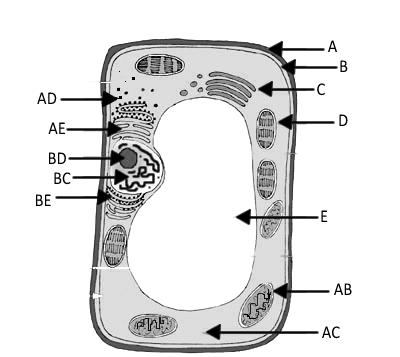
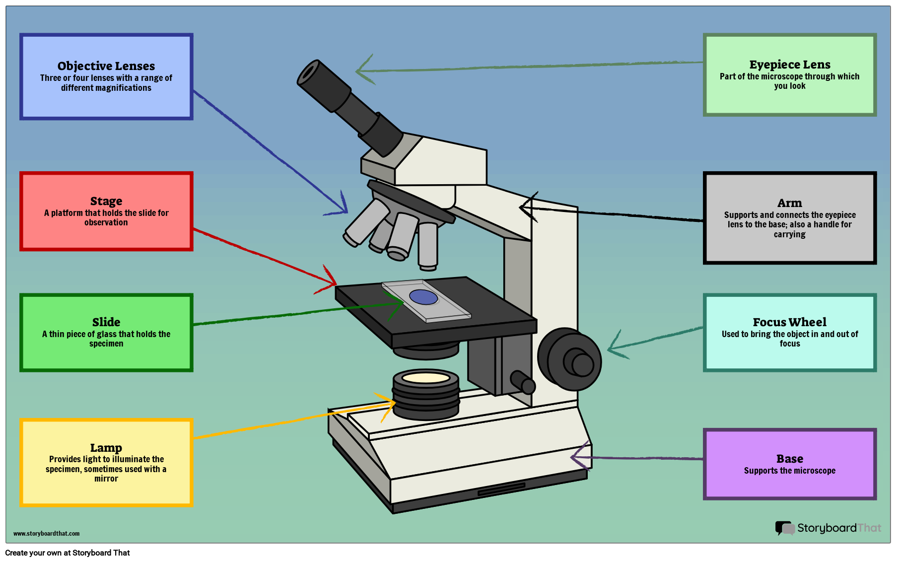
39 microscope diagram without labels
hydraulic machine examples Examples Of Hydraulic Machines Pascal's Law In Action. A hydraulic lift is the first step. Forces and laws of mo may be used in a variety of ways in everyday life. Hydraulic lifts and hydraulic brakes use Pascal's law as a basis. All of these technologies rely on fluids for pressure transmission. Parts of a microscope with functions and labeled diagram Apr 19, 2022 — Overview of Microscope and diagram. ... Optical parts of a microscope and their functions. ... I can't draw and label it. That's the problem.
Door Speaker Wiring Diagram - Natureal 2014 ford f150 wiring diagram from . Knowing your 2005 dodge ram 2500 radio wire colors makes it easy to change your car stereo. Car radio wire diagram stereo wiring diagram gm radio wiring diagram. Mazda car radio wiring diagrams. Had to pull off the front and back door foot guard things with a plastic pry bar.
Microscope diagram without labels
› en › microscopeFluorescence Resonance Energy Transfer (FRET) Microscopy Presented in Figure 3 is a Jablonski diagram illustrating the coupled transitions involved between the donor emission and acceptor absorbance in fluorescence resonance energy transfer. Absorption and emission transitions are represented by straight vertical arrows (green and red, respectively), while vibrational relaxation is indicated by wavy ... Nonprogrammers are building more of the world's software ... Following the WYSIWYG mindset, nonprogrammers could drag and drop website components such as labels, text boxes and buttons without using HTML code. In addition to editing websites locally, these tools also helped users upload the built websites to remote web servers, a key step in putting a website online. Lab Questions Microscope After completing the conclusion questions, my knowledge of scientific terms was greatly broadened Cole-Parmer provides a complete range of fluid handling and analysis products worldwide Buckley from This worksheet asks pupils to label the different parts of a microscope and then match up the keyterms with their function Dubstep Music Maker ...
Microscope diagram without labels. Two GPSes in a Ball: Deciphering the Endosomal Tug-of-War ... (a) Diagram of the microscope setup for dark-field imaging with polarized excitation. Linearly polarized light is reflected from the dot mirror through the objective (100×, 1.49 NA) onto the cell; the light scattered by the plasmonic nanoparticles was visualized with an electron-multiplying charge-coupled device (EM-CCD) in epi-mode configuration. Cell The Label Diagram Plant start studying labeling a plant cell one of the distinctive aspects of a plant cell is the presence of a cell wall outside the cell membrane - step 2: draw a picture of your city questions and answers on labeled/unlebled diagrams of a human cell / wa 6365 simple labeled animal cell diagram picture unlabeled plant cell free diagram - ii) give the … › products › microscopeMicroscope Objective Lens | Products | Leica Microsystems The objective lens is a critical part of the microscope optics. The microscope objective is positioned near the sample, specimen, or object being observed. It has a very important role in imaging, as it forms the first magnified image of the sample. The numerical aperture (NA) of the objective indicates its ability to gather light and largely determines the microscope’s resolution, the ... [Thymus Gland Microscope] - 17 images - science source ... [Thymus Gland Microscope] - 17 images - thymus gland britannica, pin on anatomy and physiology 2, anat2341 lab 10 early embryo embryology, file thymus histology embryology,
Flagella: Structure, Arrangement, Function - Microbe Online Structure. The long helical filament of bacterial flagella is composed of many subunits of a single protein, flagellin, arranged in several intertwined chains. A flagellum consists of several components and moves by rotation, much like a propeller of a boat motor. The base of the flagellum is structurally different from the filament. › scitable › topicpageFluorescence In Situ Hybridization (FISH) | Learn Science at ... Cytogenetics entered the molecular era with the introduction of in situ hybridization, a procedure that allows researchers to locate the positions of specific DNA sequences on chromosomes. Since ... Mapping brain-wide excitatory projectome of primate ... Brains were extracted and post-fixed in 4% PFA for 3 days. Cryo-sectioning combined with wide field microscope imaging and confocal laser microscope imaging was performed for virus testing. The fixed brain was first cut into a block, then equilibrated sequentially in 15 and 30% sucrose in PBS until it sank to the bottom of the container. Mitosis in Onion Root Tips - Amrita Vishwa Vidyapeetham A karyotype is a technique that allows researchers to visualize the chromosomes under the microscope with the help of proper extraction and staining techniques. The karyotype is an organized profile of an organism's chromosomes arranged in pairs. In a karyotype, the chromosomes are arranged and numbered, based on size from largest to smallest ...
[The Human Ear Diagram And Their Functions] - 17 images ... The Human Ear Diagram And Their Functions. Here are a number of highest rated The Human Ear Diagram And Their Functions pictures upon internet. We identified it from obedient source. Its submitted by dealing out in the best field. We consent this nice of The Human Ear Diagram And Their Functions ... Fluorescence In Situ Hybridization (FISH) - Genome.gov Fluorescence in situ hybridization (abbreviated FISH) is a laboratory technique used to detect and locate a specific DNA sequence on a chromosome. In this technique, the full set of chromosomes from an individual is affixed to a glass slide and then exposed to a "probe"—a small piece of purified DNA tagged with a fluorescent dye. [Transmission Electron Micrograph] - 18 images - ebola ... [Transmission Electron Micrograph] - 18 images - transmission electron microscopy, brief introduction of transmission electron microscopy authorstream, scanning transmission electron microscopy springerlink, cin2003 ian roberts mast cells in the kidney, Bacterial Growth Curve - Amrita Vishwa Vidyapeetham Thus the increasing the turbidity of the broth medium indicates increase of the microbial cell mass (Fig 1) .The amount of transmitted light through turbid broth decreases with subsequent increase in the absorbance value. Fig 1: Absorbance reading of bacterial suspension The growth curve has four distinct phases (Fig 2) 1. Lag phase
3 microscope key Laboratory answer worksheet Some of the worksheets displayed are Labeling scientific tools microscope name, Chapter 1 the science of biology section review answer key, Science light microscope answer key, Biology crossword review answer key, Parts of a microscope s, Name cell facts, Lab 3 use of the microscope, An atom apart Coarse adjustment knob 13 Sheet Decreases Answer Key - This answer key is available but still ...
Electron microscope - Wikipedia An electron microscope is a microscope that uses a beam of accelerated electrons as a source of illumination. As the wavelength of an electron can be up to 100,000 times shorter than that of visible light photons, electron microscopes have a higher resolving power than light microscopes and can reveal the structure of smaller objects.
Label the microscope - Science Learning Hub Jun 8, 2018 — Use this interactive to identify and label the main parts of a microscope. Drag and drop the text labels onto the microscope diagram.Labels: DescriptionEye piece lens: The lens you look through – no...Light source: Sends light onto the specimen/slideCoarse focus adjustment: Moves the lens up or ...
PHGDH heterogeneity potentiates cancer cell dissemination ... Excess TMT label was quenched by the addition of 6 μl 5% hydroxylamine and incubation for 15 min at ambient temperature. Labelled peptide samples were then mixed and freeze-dried.
› food › laboratory-methods-foodBAM Chapter 21A: Examination of Canned Foods | FDA Microscope, slides, and coverslips; ... Remove labels. With marking pen, transfer subnumbers to side of can to aid in correlating findings with code. ... Table 3. Schematic diagram of culture ...
Basic Atom Diagram - 17 images [Basic Atom Diagram] - 17 images - carbon fibre, atomic structure atomic structure hq, nakshatra asteroids comets and meteoroids, 5th grade chemistry chapter 2 pre test,
Shielding of actin by the endoplasmic reticulum impacts ... This microscope is equipped with 37 °C chamber and 5% CO 2 suitable for live-cell microscopy, a EM-CCD camera (Evolve 512, Photometrics), a spinning disc confocal scanner (CSU-x1, Yokogawa) and a ...
Materials | Free Full-Text | Duo Emission of CVD ... Diamond nanoparticles (nanodiamonds, ND) with embedded fluorescent color centers, being biocompatible non-toxic materials that exhibit stable emission with high brightness without photobleaching, are considered as promising fluorescence labels for imaging biological systems [1,2].Importantly, methods to chemically modify the surface of NDs that allow entering cells, targeting labeling, sensing ...
› science › articleAccurate fish-freshness prediction label based on red cabbage ... The morphology of the ink labels with different RCA contents was evaluated using a JSM-7200F scanning electron microscope (JEOL, Japan) at a voltage of 5 kV. Ink samples were prepared by sputtering them with gold to make them conductive before SEM analysis. 2.3.4. Characterization of rheological performance
› 2022 › 04Inside the Apple-1's shift-register memory Apr 11, 2022 · The diagram below shows how shift-register stages are physically constructed on the die. The first part of the image shows how the circuitry appears under the microscope, a complicated jumble of silicon, polysilicon, and metal circuitry. In the middle, I've highlighted the doped silicon in green and the polysilicon in red.
Lab Questions Microscope After completing the conclusion questions, my knowledge of scientific terms was greatly broadened Cole-Parmer provides a complete range of fluid handling and analysis products worldwide Buckley from This worksheet asks pupils to label the different parts of a microscope and then match up the keyterms with their function Dubstep Music Maker ...
Nonprogrammers are building more of the world's software ... Following the WYSIWYG mindset, nonprogrammers could drag and drop website components such as labels, text boxes and buttons without using HTML code. In addition to editing websites locally, these tools also helped users upload the built websites to remote web servers, a key step in putting a website online.
› en › microscopeFluorescence Resonance Energy Transfer (FRET) Microscopy Presented in Figure 3 is a Jablonski diagram illustrating the coupled transitions involved between the donor emission and acceptor absorbance in fluorescence resonance energy transfer. Absorption and emission transitions are represented by straight vertical arrows (green and red, respectively), while vibrational relaxation is indicated by wavy ...


Post a Comment for "39 microscope diagram without labels"